Foot Muscles Mri : 1 : These muscles begin and attach within the skeleton of the foot, have complex anatomical and topographical and functional relationships with.
byAdmin•
0
Foot Muscles Mri : 1 : These muscles begin and attach within the skeleton of the foot, have complex anatomical and topographical and functional relationships with.. What to look for, where to look & how to report. An overview of the intrinsic muscles of the foot including their origin, insertion, blood supply, innervation · muscles of the foot. Posted by radiologyer at 8:12 am. Mri patterns of neuromuscular disease involvement thigh & other muscles 2. Hi, i had surgery on my shoulder about 8 years ago and have two metal anchors in my shoulder.
Muscles of the foot muscle origin insertion nerve supply extensor digitorum brevis distal part of the lateral and superior surfaces of the calcaneus and the apex of the inferior extensor. Intrinsic muscles of the feet part 2 | layers 3 & 4. Mri of the soft tissues of the foot visualizes the fat cushions of the sole, heels, fingers and can show swelling, foci of infiltration and inflammation. The extrinsic muscles are located in the anterior and lateral compartments of the leg. Posted by radiologyer at 8:12 am.
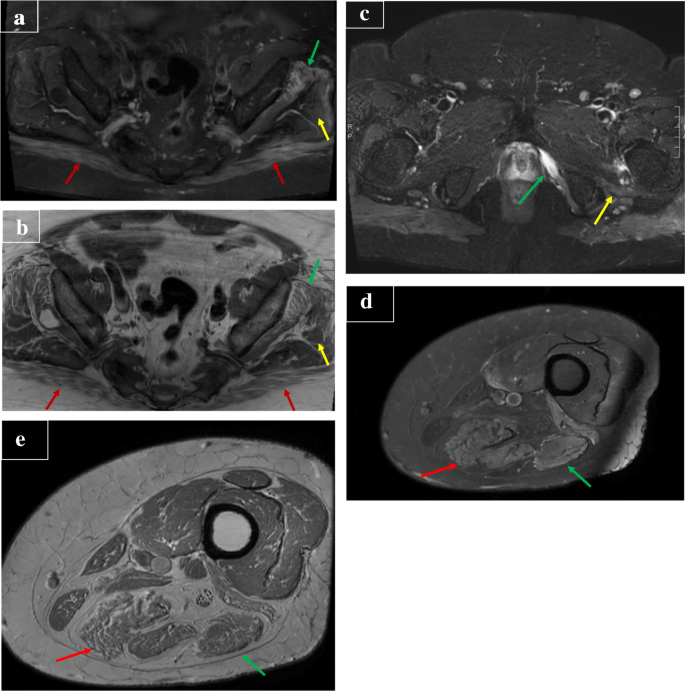
Covid 19 Related Muscle Denervation Atrophy Springerlink from media.springernature.com Muscle mri sequences & patterns asymmetric myopathy hereditary acquired connective tissue neurogenic. Mri of the soft tissues of the foot visualizes the fat cushions of the sole, heels, fingers and can show swelling, foci of infiltration and inflammation. Indications for foot mri scan. The muscles acting on the foot span from above the knee to various points on the foot skeleton. Intrinsic muscles of the feet part 2 | layers 3 & 4. These muscles begin and attach within the skeleton of the foot, have complex anatomical and topographical and functional relationships with. By muhammad ali, mb bs; The extrinsic muscles are located in the anterior and lateral compartments of the leg.
Lateral and medial processes of calcaneal tuberosity.
The flexor digiti minimi brevis (flexor brevis minimi digiti, flexor digiti quinti brevis) lies under the metatarsal bone on the little toe, and resembles one of the interossei. Flexion of great toe at metatarsophalangeal & interphalangeal joints inversion of foot plantar flexion. Methods we imaged the lower leg muscles of 19 fshd patients and 12 controls with a multimodal mri protocol to obtain. Mri with hardware in foot? Indications for foot mri scan. Muscles of the ankle and foot. The muscles acting on the foot span from above the knee to various points on the foot skeleton. ► hip ► pelvis ► thigh ► knee ► lower extremity/shin ► ankle ► foot. Intrinsic muscles of the feet part 2 | layers 3 & 4. Thank you for your attention. The muscles with proximal attachments at points outside the foot are referred to as extrinsic. A magnetic resonance imaging (mri) was performed on a normal subject; Learn about foot and ankle mri here.
Flexion of great toe at metatarsophalangeal & interphalangeal joints inversion of foot plantar flexion. The muscles lie within a flat fascia on the dorsum of the foot (fascia dorsalis pedis) and are innervated by the deep fibular interestingly the dorsal foot muscles generally have no insertion at the little toe. 12 photos of the foot muscle anatomy mri. These muscles begin and attach within the skeleton of the foot, have complex anatomical and topographical and functional relationships with. Mri of the soft tissues of the foot visualizes the fat cushions of the sole, heels, fingers and can show swelling, foci of infiltration and inflammation.
Characteristic Mri Findings Of Epidermal Cysts Categorized By Size Fulltext from benthamopen.com Bone contusions, osteonecrosis, marrow oedema syndromes, and stress > fractures) > synovial based disorders ( e.g. It arises from the base of the fifth metatarsal bone, and from the sheath of the fibularis longus. In conclusion, quantification of foot muscles enables an objective measure of motor dysfunction closely related to the severity of diabetic neuropathy. Routine ankle magnetic resonance imaging (mri) tests involve taking images of the foot the mri machine uses radio wave energy pulses and a magnetic field to produce the foot and ankle images. Intrinsic muscles of the feet part 2 | layers 3 & 4. Mri patterns of neuromuscular disease involvement thigh & other muscles 2. Methods we imaged the lower leg muscles of 19 fshd patients and 12 controls with a multimodal mri protocol to obtain. ► hip ► pelvis ► thigh ► knee ► lower extremity/shin ► ankle ► foot.
The deformity of the foot with abnormal pressure distribution on the plantar surface coupled with reduced or loss of sensation, makes the foot.
The muscles acting on the foot can be divided into two distinct groups; Indications for foot mri scan. Upper and lower lines mark. A magnetic resonance imaging (mri) was performed on a normal subject; What to look for, where to look & how to report. The flexor digiti minimi brevis (flexor brevis minimi digiti, flexor digiti quinti brevis) lies under the metatarsal bone on the little toe, and resembles one of the interossei. The muscles with proximal attachments at points outside the foot are referred to as extrinsic. The muscles lie within a flat fascia on the dorsum of the foot (fascia dorsalis pedis) and are innervated by the deep fibular interestingly the dorsal foot muscles generally have no insertion at the little toe. Bone contusions, osteonecrosis, marrow oedema syndromes, and stress > fractures) > synovial based disorders ( e.g. Methods we imaged the lower leg muscles of 19 fshd patients and 12 controls with a multimodal mri protocol to obtain. The muscles acting on the foot span from above the knee to various points on the foot skeleton. Posted by radiologyer at 8:12 am. ► hip ► pelvis ► thigh ► knee ► lower extremity/shin ► ankle ► foot.
Indications for foot mri scan. What to look for, where to look & how to report. The muscles acting on the foot can be divided into two distinct groups; Lateral and medial processes of calcaneal tuberosity. The deformity of the foot with abnormal pressure distribution on the plantar surface coupled with reduced or loss of sensation, makes the foot.
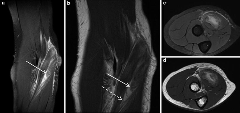
Mri Of Muscle Injuries Springerlink from media.springernature.com Mri with hardware in foot? Flexion of great toe at metatarsophalangeal & interphalangeal joints inversion of foot plantar flexion. Gray's anatomy for students, 2nd ed. Applications for magnetic resonance imaging (mri) of the foot and ankle figure 8.4 image planes for foot and ankle mri. The muscles acting on the foot can be divided into two distinct groups; Thank you for your attention. If you'd like to support us and get something great in return. Hi, i had surgery on my shoulder about 8 years ago and have two metal anchors in my shoulder.
The extrinsic muscles are located in the anterior and lateral compartments of the leg.
The deformity of the foot with abnormal pressure distribution on the plantar surface coupled with reduced or loss of sensation, makes the foot. Muscles of the ankle and foot. Intrinsic muscles of the feet part 2 | layers 3 & 4. Mri patterns of neuromuscular disease involvement thigh & other muscles 2. The muscles acting on the foot can be divided into two distinct groups; The extrinsic muscles are located in the anterior and lateral compartments of the leg. Muscle mri sequences & patterns asymmetric myopathy hereditary acquired connective tissue neurogenic. By muhammad ali, mb bs; Mri of the soft tissues of the foot visualizes the fat cushions of the sole, heels, fingers and can show swelling, foci of infiltration and inflammation. If you'd like to support us and get something great in return. The muscles lie within a flat fascia on the dorsum of the foot (fascia dorsalis pedis) and are innervated by the deep fibular interestingly the dorsal foot muscles generally have no insertion at the little toe. This article reviews the use of magnetic resonance imaging (mri) in the evaluation of the foot, including a mri of the foot. Indications for foot mri scan.